
Special X-Ray Procedures: Advancing Diagnostic Precision in Modern Healthcare
- Admin

- Mar 26
- 4 min read
X-ray imaging is the cornerstone of medical diagnostics. Routine X-rays are effectively used for detecting fractures and basic abnormalities but modern medicine increasingly relies on special X-ray procedures for deeper and more precise evaluation.
These advanced techniques combine contrast agents, real-time imaging, and minimally invasive interventions to provide highly detailed insights into the body. From diagnosing complex conditions to guiding treatments, special X-ray procedures play a crucial role in improving patient outcomes.
What Are Special X-Ray Procedures?
Special X-ray procedures are advanced imaging techniques that extend beyond routine radiography to offer detailed views of internal body structures. By using contrast agents, real-time imaging, and image-guided techniques, they support accurate diagnosis and treatment of various medical conditions. These procedures enhance precision, enable minimally invasive care, and play a crucial role in modern healthcare.
This process typically involve:
Contrast media (like Barium, Iodine)
Real-time imaging (Fluoroscopy)
Image-guided interventions
Together, these methods allow doctors to clearly visualize soft tissues, blood vessels, and organ function, providing far more detailed information than conventional X-rays.
1. Contrast-Based X-Ray Studies
One of the most widely used categories of special X-ray procedures involves contrast media, which helps highlight specific organs or systems. Contrast studies enhance visibility of internal organs by introducing a radiopaque substance. This is especially useful for detecting blockages, ulcers, or structural abnormalities.
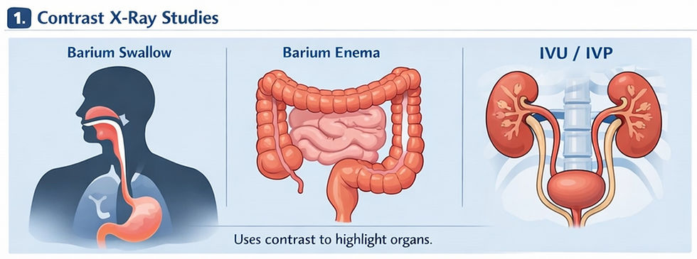
Common Examples:
Barium Swallow / Barium Meal: Used to examine the esophagus, stomach, and upper gastrointestinal tract.
Barium Enema: Helps evaluate the large intestine for abnormalities like polyps or tumors.
Intravenous Urography (IVU/IVP): Assesses the kidneys, ureters, and bladder by injecting contrast into a vein.
2. Fluoroscopy (Real-Time Imaging)
Fluoroscopy is a real-time X-ray imaging technique that produces continuous moving images of internal structures. It is widely used to study organ function, guide medical procedures, and assist in interventions like catheter placement, joint examinations, and gastrointestinal studies with enhanced diagnostic accuracy. Fluoroscopy plays a crucial role in both diagnosis and treatment, particularly when precision and real-time monitoring are required.

Key Applications:
Swallowing studies to detect aspiration or motility issues
Orthopedic assessments of joint movement
Guidance during catheter insertions and other procedures
3. Angiography (Blood Vessel Imaging)
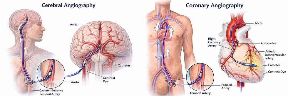
Angiography is a specialized imaging procedure used to visualize blood vessels using contrast dye and X-rays. It helps detect blockages, narrowing, or abnormalities in arteries and veins. Commonly used for heart, brain, and peripheral vessels, angiography also guides interventional procedures like angioplasty and stent placement for effective treatment.
Types of Angiography:
Coronary Angiography: Evaluates blood flow in the heart’s arteries
Cerebral Angiography: Examines blood vessels in the brain
Peripheral Angiography: Assesses circulation in the limbs
4. Mammography

Mammography is a specialized X-ray imaging technique used to examine breast tissue for early detection of abnormalities, particularly breast cancer. It uses low-dose radiation and gentle compression to produce clear, detailed images of the breast. Mammography can identify small tumors, calcifications, and other changes before symptoms appear. Regular screening significantly improves early diagnosis, enabling timely treatment, better outcomes, and increased survival rates among women.
5. Interventional Radiology
Interventional Radiology is a medical specialty that uses imaging techniques like X-ray and ultrasound to perform minimally invasive procedures. It helps diagnose and treat conditions such as blocked arteries, tumors, and internal bleeding. These procedures reduce the need for surgery, resulting in faster recovery, less pain, and lower risk for patients.
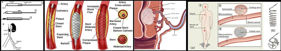
Common Procedures:
Angioplasty and stent placement
Biopsies
Drainage of abscesses
Tumor embolization
6. Myelography

Myelography is a specialized imaging procedure that uses contrast dye and X-rays to examine the spinal cord, nerve roots, and spinal canal. It helps diagnose conditions such as herniated discs, spinal stenosis, and tumors. This technique is especially useful when MRI is not suitable or when detailed spinal imaging is required.
It is particularly useful for detecting:
Herniated discs
Spinal stenosis
Tumors or nerve compression
7. Hysterosalpingography (HSG)
Hysterosalpingography (HSG) is a specialized X-ray procedure used to study the uterus and fallopian tubes to evaluate female firtility. A contrast dye is injected to check for blockages or abnormalities. It is commonly used in infertility investigations, helping doctors assess tubal patency and uterine structure, and guiding further treatment or reproductive planning. A contrast agent is introduced into the uterus through the vagina to identify the following:
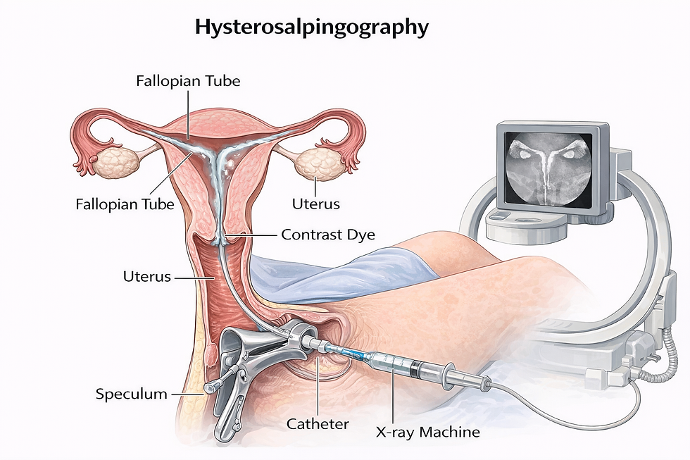
• Blocked fallopian tubes
• Uterine abnormalities
• Ovarian cysts
• Causes of infertility
8. Arthrography
Arthrography is a specialized imaging procedure where contrast dye is injected into a joint to visualize structures like ligaments, cartilage, and joint capsules. It helps diagnose tears, instability, and joint disorders, often using X-ray or fluoroscopy for accurate and detailed assessment. This is very useful in sports injuries and ligament damage.
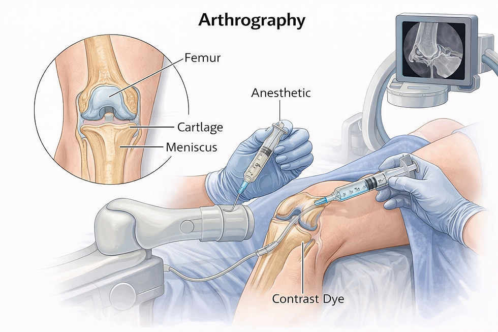
It is commonly used for:
Shoulder injuries
Knee ligament tears
Joint instability
9. Dental & Bone Density Imaging
Dental and bone density imaging use specialized X-ray techniques to assess oral health and bone strength. Dental X-rays detect cavities and jaw issues, while DEXA scans measure bone mineral density, helping diagnose osteoporosis and monitor fracture risk for better long-term skeletal health.
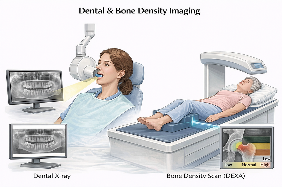
This process includes the following:
OPG & Cephalometric X-rays provides panoramic and skeletal views for dental and orthodontic assessment.
DEXA Scan measures bone mineral density, helping diagnose osteoporosis and assess fracture risk accurately.
Advantages of Special X-Ray Procedures
Special X-ray procedures offer advanced imaging capabilities that improve diagnostic accuracy and patient care. By combining enhanced visualization, real-time assessment, and minimally invasive techniques, these methods enable faster detection and effective treatment of various medical conditions, improving overall clinical outcomes.
Enhanced visualization of soft tissues
Real-time functional assessment
Minimally invasive techniques
Faster and more accurate diagnosis
Contrast Media and Its Effects
Contrast media plays a key role in these procedures by highlighting internal structures. However, in some cases, it may cause reactions, therefore, it is mandatory to obtain patient consent before performing any contrast-based procedure.:
• Mild: Nausea, vomiting, skin rash (urticaria)
• Moderate: Mild breathing difficulty, increased heart rate, redness
• Severe: Loss of consciousness, severe bronchospasm
Emergency Drugs in Special Procedures
Since contrast media may sometimes cause allergic or severe reactions, emergency drugs must always be readily available. These medications are essential for immediate management of complications:
• Allergy: Avil, Decadron
• Pain relief: Paracetamol
• Cardiac support: Aspirin, Hydrocortisone
• Diabetes emergency: Glucagon
• Respiratory support: Salbutamol
Conclusion
Special X-ray procedures have transformed modern diagnostic imaging by offering deeper insights and enabling targeted treatments. From contrast studies to interventional radiology, these techniques are indispensable in today’s healthcare system.
As technology continues to advance, these procedures will become even more precise, accessible, and patient-friendly—further improving diagnostic accuracy and treatment outcomes.


Comments